Bone Cross Section Histology / Pin by Danielle Papas on Nursing Anatomy & Physiology ... / Cross and longitudinal sections (unstained).
byAdmin-
0
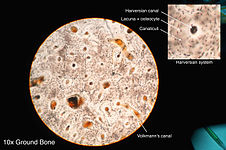
Bone Cross Section Histology / Pin by Danielle Papas on Nursing Anatomy & Physiology ... / Cross and longitudinal sections (unstained).. Histology of the haversian system (osteons, lamellae, canaliculi, volkmann's canals, and circumferential lamellae) in a ground bone section. A cross section of a typical osteon or haversian system. Note that in tubal cross sections, circular smooth muscle layers will have a longitudinal section while longitudinal layers will be in cross section. Cross and longitudinal sections (unstained). In addition to discussing the cellular constituents of bone and the architectural arrangement of their products.
The histology of compact bone. A cross section of any bone will demonstrate these two types of bones. A cross section of a typical osteon or haversian system. Filopodia from adjacent osteocytes communicate via gap junctions. Histology classification of bone tissue.
Osteon - Wikipedia from upload.wikimedia.org Learn vocabulary, terms and more with flashcards, games and other study tools. A cross section of a human long bone. From wikimedia commons, the free media repository. The histology of compact bone. Cross section of a long bone. In development there are 2 separate signaling pathways for pattern formation and the formation of bone itself. The literature on juvenile cortical bone histology is. 'compact or cortical bone is usually thick dense bone that forms the outer shell cross sections of the bone when studied under the microscope reveal quite a different picture.
Since the denser compact bone.
Note that in tubal cross sections, circular smooth muscle layers will have a longitudinal section while longitudinal layers will be in cross section. This image shows compact bone in cross section. Histology of bone gross structure • the diaphysis is the shaft and notably comprises the marrow bone tissues • the medullary cavity comprises spongy bone. Cross and longitudinal sections (unstained). A cross section of a typical osteon or haversian system. The central macrophage is often difficult to identify in histologic sections. The term 'bone marrow' (bm) refers to the tissue occupying the cavities under the cortex within the this chapter will describe the histology of bm in the trephine biopsy. (gross and histology) aimi nadia razlan centre of preclinical science studies fa c u lt y o f d e n t i s t ry 01 02 03 describe the describe the describe types of bones with regards histology of bone ossification to its classification and. Histology hint from sarah bellham: The significance of histological examination in the classification and diagnosis of clinical conditions is reliant on the expertise of the histology laboratory in managing the wide spectrum of specimen types submitted for analysis. In these sections, the trapped air bends the light giving a dark image; Filopodia from adjacent osteocytes communicate via gap junctions. The histology of compact bone.
The significance of histological examination in the classification and diagnosis of clinical conditions is reliant on the expertise of the histology laboratory in managing the wide spectrum of specimen types submitted for analysis. Lamellar bone forms both trabecular bone and compact bone, which are the two macroscopically recognizable bone forms. A cross section of a human long bone. Jump to navigation jump to search. The literature on juvenile cortical bone histology is.
Respiratory - Human Anatomy Histology with Several at Lake ... from classconnection.s3.amazonaws.com The literature on juvenile cortical bone histology is. (gross and histology) aimi nadia razlan centre of preclinical science studies fa c u lt y o f d e n t i s t ry 01 02 03 describe the describe the describe types of bones with regards histology of bone ossification to its classification and. Bone decalcification is the removal of the mineral component using an acid, leaving the bone soft and easy to cut. Jump to navigation jump to search. First, let's look at a section of. Histology classification of bone tissue. The central macrophage is often difficult to identify in histologic sections. • the outer layers of the diaphysis now, let's point out these histological structures in bone specimens.
Histology hint from sarah bellham:
Lamellar bone forms both trabecular bone and compact bone, which are the two macroscopically recognizable bone forms. What follows is primarily a guide to observing particular features microscopically. The section may be either cross section (x.s.) or longitudinal section (l.s.). First, study cross sections (slides 51 and 93b). From wikimedia commons, the free media repository. Both sections have been decalcified in order to make it easier to cut the bone into thin sections, but the collagen is still present in the slides. Available at the itunes store and for android users at the google play store. The literature on juvenile cortical bone histology is. 'compact or cortical bone is usually thick dense bone that forms the outer shell cross sections of the bone when studied under the microscope reveal quite a different picture. The histology of compact bone. Bone decalcification is the removal of the mineral component using an acid, leaving the bone soft and easy to cut. Dry bone is cut and polished before mounting on a slide. Contents (click on desired chapter).
Learn vocabulary, terms and more with flashcards, games and other study tools. A cross section of any bone will demonstrate these two types of bones. Bone decalcification is the removal of the mineral component using an acid, leaving the bone soft and easy to cut. *blood vessels *nerves *loose connective tissue. This is a cross section through decalcified bone.
Bone Histology - Biology bibliographies - Cite This For Me from mesa-anatomy.weebly.com Lamellar bone forms both trabecular bone and compact bone, which are the two macroscopically recognizable bone forms. What follows is primarily a guide to observing particular features microscopically. Since the denser compact bone. Bone decalcification is the removal of the mineral component using an acid, leaving the bone soft and easy to cut. Available at the itunes store and for android users at the google play store. Histology classification of bone tissue. The literature on juvenile cortical bone histology is. Histology hint from sarah bellham:
Dry bone is cut and polished before mounting on a slide.
Use the illustrations in your textbook as a guide and identify with the scanning objective the following structures. Histology hint from sarah bellham: First, let's look at a section of. Contents (click on desired chapter). In development there are 2 separate signaling pathways for pattern formation and the formation of bone itself. Anyway, examine the fibers cut in xs to see that the nuclei are located in the center of the fibers (you may need to use oil emersion). Histology of the haversian system (osteons, lamellae, canaliculi, volkmann's canals, and circumferential lamellae) in a ground bone section. First, study cross sections (slides 51 and 93b). By and large they could be either mineralised or. The central macrophage is often difficult to identify in histologic sections. Cross and longitudinal sections (unstained). (gross and histology) aimi nadia razlan centre of preclinical science studies fa c u lt y o f d e n t i s t ry 01 02 03 describe the describe the describe types of bones with regards histology of bone ossification to its classification and. The histology of compact bone.
Haversian systems (osteons) are distinctive structural units of compact bone that reflect the developmental and nutritive pattern of its lamellar bone cross section. A cross section of a human long bone.